Lower Back Muscles Labeled - Axial Muscles Of The Head Neck And Back Anatomy Physiology : These sections are cervical (neck), thoracic (upper and middle back), lumbar (lower back), and sacrum (tailbone).
Lower Back Muscles Labeled - Axial Muscles Of The Head Neck And Back Anatomy Physiology : These sections are cervical (neck), thoracic (upper and middle back), lumbar (lower back), and sacrum (tailbone).. They help to bend the back to one side or the other. Muscles of lower back diagram in this image, you will find an occipital bone, sternocleidomastoid, trapezius, deltoid in muscles of the lower back diagram. Exercise of this organ system is critical to prevent wasting from age or th… Human musculature bodybuilding infographic muscular system vector human anatomy back muscle anatomy bicep male muscular anatomy human body anatomy female female anatomy muscle hamstrings muscle. See more ideas about massage therapy, back pain, muscle anatomy.
See more ideas about massage therapy, back pain, muscle anatomy. Related posts of muscles of the lower back and buttocks diagram smooth muscle diagram labeled. Lower back muscles labeled educational anatomical scheme vector illustration. The vertebral column of the lower back includes the five lumbar vertebrae, the sacrum, and the coccyx. It is often referred to as a pulled muscle.
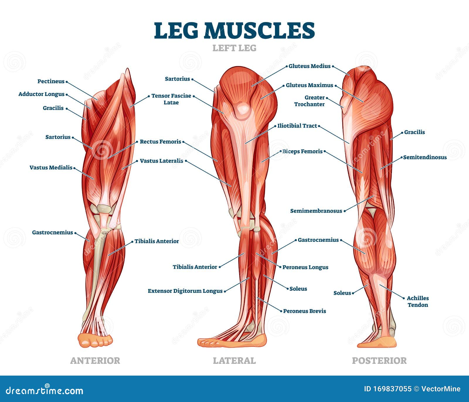
Leg Muscle Stock Illustrations 8 717 Leg Muscle Stock Illustrations Vectors Clipart Dreamstime from thumbs.dreamstime.com As you can see, there are also have a spine of scapula deltoid, triceps brachii, latissimus dorsi. This website uses cookies to improve your experience while you navigate through the website. Lower back strain is acute pain caused by damage to the muscles and ligaments of the back. Only the iliocostalis is shown on the right side. These sections are cervical (neck), thoracic (upper and middle back), lumbar (lower back), and sacrum (tailbone). The muscle then courses up to your shoulder and attaches to your upper arm bone. The lumbar and sacrum region make up the bone of the lower back anatomy. This curve, called lordosis, helps to:
The lumbar spine is the lower back that begins below the last thoracic vertebra (t12) and ends at the top of the sacral spine, or sacrum (s1).
The bones of the pelvis and lower back work together to support the body's weight, anchor the abdominal and hip muscles, and protect the delicate vital organs of the vertebral and abdominopelvic cavities. The muscles of the lower back, including the erector spinae and quadratus lumborum muscles, contract to extend and laterally bend the vertebral column. Muscles of the lower back and hip diagram, human muscles, muscles of the lower back and hip diagram. Lumbar muscle strain is caused when muscle fibers are abnormally stretched or torn. Deep muscles of the lower back include: The multifidus, a long muscle that travels nearly the entire length of the back.it helps to stabilize and rotate the lower back, and additionally takes some. The erector spinae is composed of three subgroups: See back muscle anatomy stock video clips. Related posts of muscles of the lower back and buttocks diagram smooth muscle diagram labeled. These sections are cervical (neck), thoracic (upper and middle back), lumbar (lower back), and sacrum (tailbone). This is a tutorial to quickly s. Extrinsic and intrinsic.the back functions are many, such as to house and protect the spinal cord, hold the body and head upright, and adjust the movements of the upper and lower limbs. The spine's four sections, from top to bottom, are the cervical (neck), thoracic (abdomen,) lumbar (lower back), and sacral (toward tailbone).
Posterior view of the erector spinae musculature of the low back. It comprises the vertebral column (spine) and two compartments of back muscles; The muscle then courses up to your shoulder and attaches to your upper arm bone. Each lumbar spinal level is numbered from top to bottom—l1 through l5, or l6. Extends and laterally bends the neck and head, rotates head to the same side:
Human Anatomy And Physiology Of Muscles Human Muscle Anatomy Muscle Anatomy Muscle Diagram from i.pinimg.com Related posts of muscles of the lower back and buttocks diagram smooth muscle diagram labeled. Posterior view of the erector spinae musculature of the low back. Your lats are a major back muscle and mover of your shoulder joint. This website uses cookies to improve your experience while you navigate through the website. The lumbar spine is the lower back that begins below the last thoracic vertebra (t12) and ends at the top of the sacral spine, or sacrum (s1). Throughout the spine, intervertebral discs made of. See more ideas about massage therapy, back pain, muscle anatomy. This curve, called lordosis, helps to:
Related posts of muscles of the lower back and buttocks diagram smooth muscle diagram labeled.
Muscles of the lower back and hip diagram, human muscles, muscles of the lower back and hip diagram. The muscles that move the upper legs (thigh) there are many muscles that move the large bone of the thigh. The spine's four sections, from top to bottom, are the cervical (neck), thoracic (abdomen,) lumbar (lower back), and sacral (toward tailbone). This website uses cookies to improve your experience while you navigate through the website. Illustration about labeled, drawing, back, human, diagram, oblique, medical, anatomy. These sections are cervical (neck), thoracic (upper and middle back), lumbar (lower back), and sacrum (tailbone). By the way, have you heard about the myth of. Here is a depiction of the skeletal frame with the lower. The muscles of the lower back, including the erector spinae and quadratus lumborum muscles, contract to extend and laterally bend the vertebral column. Muscle origin insertion action innervation artery notes; See more ideas about massage therapy, back pain, muscle anatomy. Out of these, the cookies that are categorized as necessary are stored on your browser as they are essential for the working of basic functionalities of the website. The erector spinae is composed of three subgroups:
The lumbar and sacrum region make up the bone of the lower back anatomy. Only the iliocostalis is shown on the right side. The muscles of the lower back, including the erector spinae and quadratus lumborum muscles, contract to extend and laterally bend the vertebral column. Bones of the pelvis and lower back. Extrinsic and intrinsic.the back functions are many, such as to house and protect the spinal cord, hold the body and head upright, and adjust the movements of the upper and lower limbs.
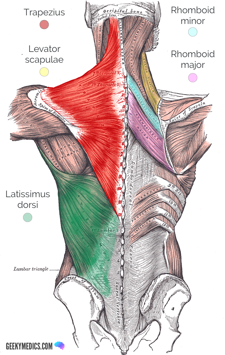
Superficial Back Muscles Anatomy Geeky Medics from geekymedics.com The quadratus lumborum muscles (orange, in the image above) are found in the lower back (also called the lumbar area). All three subgroups are shown on the left side; The back is the body region between the neck and the gluteal regions. These muscles provide posture and stability to the body by holding the vertebral column erect and adjusting the position of the body to maintain balance. Related posts of muscles of the lower back and buttocks diagram smooth muscle diagram labeled. Your lats are a major back muscle and mover of your shoulder joint. Mastoid process and lateral end of the superior nuchal line: The multifidus, a long muscle that travels nearly the entire length of the back.it helps to stabilize and rotate the lower back, and additionally takes some.
Throughout the spine, intervertebral discs made of.
Muscle strains and sprains are common in the lower back, because it supports the weight of the upper body and is involved in moving, twisting and bending. See more ideas about massage therapy, back pain, muscle anatomy. It also covers some common conditions and injuries that can affect the back. They help to bend the back to one side or the other. Muscles of the lower back and hip diagram, human muscles, muscles of the lower back and hip diagram. The back is the body region between the neck and the gluteal regions. Extrinsic and intrinsic.the back functions are many, such as to house and protect the spinal cord, hold the body and head upright, and adjust the movements of the upper and lower limbs. This is a tutorial to quickly s. This curve, called lordosis, helps to: Lumbar (lower back) muscle strains and sprains are the most common causes of low back pain. Lower back muscles labeled educational anatomical scheme vector illustration. The quadratus lumborum muscles (orange, in the image above) are found in the lower back (also called the lumbar area). As you can see, there are also have a spine of scapula deltoid, triceps brachii, latissimus dorsi.


0 Komentar